Newcastle disease is caused by an RNA virus,
Newcastle disease virus (NDV), synonymous with
avian paramyxovirus-1 which is in
the genus Avulavirus , family Paramyxoviridae. Isolates
are
classified into 1 of 3 virulence groups by chicken embryo and chicken
inoculation as virulent
(velogenic), moderately virulent (mesogenic), or of low
virulence (lentogenic). Lentogenic strains
are used widely as live vaccines in
healthy chickens. Clinical manifestations vary from high morbidity
and mortality
to asymptomatic infections. The severity of an infection is dependent on virus
virulence
and the age, immune status, and susceptibility of the host species.
Chickens are the most and waterfowl
the least susceptible of domestic
poultry.
| Clinical Findings: |
Onset is rapid, and signs appear throughout the
flock within 2-12 days (average 5) after aerosol
exposure. Spread is slower if
the fecal-oral route is the primary means of transmission, particularly
for
caged birds. Young birds are the most susceptible. Observed signs depend on
whether the
infecting virus has a predilection for respiratory, digestive, or
nervous systems. Respiratory signs
of gasping, coughing, sneezing, and rales
predominate in low virulence infections. Nervous signs of
tremors, paralyzed
wings and legs, twisted necks, circling, clonic spasms, and complete paralysis
may
accompany, but usually follow, the respiratory signs in neurotropic
velogenic disease. Nervous signs
with diarrhea are typical in pigeons, and
nervous signs are frequently seen in cormorants and exotic
bird species.
Respiratory signs with depression, watery-greenish diarrhea, and swelling of the
tissues
of the head and neck are typical of the most virulent form of the
disease, viscerotropic velogenic
Newcastle disease (VVND, also called exotic
Newcastle disease), although nervous signs may also
be seen. Varying degrees of
depression and inappetence are observed. A partial or complete cessation
of egg
production may occur. Eggs may be abnormal in color, shape, or surface, and have
watery
albumen. Mortality is variable but can be as high as
100%.
| |
 | Description:
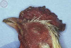
Avian, skin. There is marked hemorrhage of the comb and head, with cyanosis of the margin of the comb.
Credit: California Animal Health and Food Safety Laboratory System
Photo ID: ND_003
0205 |
|
Lesions:
Remarkable gross lesions are usually observed only
with VVND. Petechiae may be seen on the serous
membranes; hemorrhages of the
proventricular mucosa and intestinal serosa are accompanied by
multifocal,
necrotic hemorrhagic areas on the mucosal surface of the intestine, especially
at lymphoid
foci such as cecal tonsils. Splenic necrosis and hemorrhage and
edema around the thymus may also be
observed. In contrast, the lesions in birds
infected with lower virulence NDV strains may be limited
to congestion and
mucoid exudates seen in the respiratory tract with opacity and thickening of the
air
sacs. Secondary bacterial infections will increase the severity of the
respiratory lesions.
| Diagnosis: |
Diagnosis may be confirmed by isolation of a
hemagglutinating virus identified by inhibition with Newcastle disease
antiserum. A rise in hemagglutination-inhibition antibodies in paired serum
samples also confirms the disease. The acute form should be differentiated from
highly pathogenic avian influenza (Avian Influenza:
Introduction). Virulence of an isolate is established by the rapidity of
killing day-old chicks inoculated by the intracerebral route, the intracerebral
pathogenicity index, or by the presence of a specified amino acid motif at the
cleavage site of the fusion protein (F) precursor (FO). Reference laboratories
use monoclonal antibodies to detect antigenic differences and nucleotide
sequence analysis to detect genetic differences for comparison of isolates from
different outbreaks and to identify the source of those infections.
| |
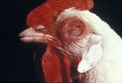
 | Description:
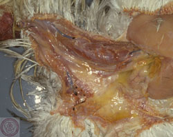
Avian, skin. There is marked subcutaneous edema in the neck, extending to the thoracic inlet.
Credit: California Animal Health and Food Safety Laboratory System |
|
|
<>
<>
<>
<>
<>
Live lentogenic vaccines, chiefly B1 and LaSota
strains, are widely used and typically administered to poultry by mass
application in drinking water or by spray. Alternatively, individual
administration is via the nares or conjunctival sac. Healthy chicks are
vaccinated as early as day 1-4 of life. However, delaying vaccination until the
second or third week avoids maternal antibody interference with an active immune
response. Mycoplasma , some other bacteria, and other
viruses affecting the respiratory tract, if present, may act synergistically
with some vaccines to aggravate the vaccine reaction after spray administration.
|
| Oil-adjuvanted inactivated vaccines are also used
following live vaccine in breeders and layers and may be used alone in
situations where use of live virus may be contraindicated. In countries where
virulent NDV is endemic, a combination of live virus and inactivated vaccine can
be used; or alternatively, if permitted by law, a live mesogenic strain vaccine
may be used in older birds. The frequency of revaccination to protect chickens
throughout life largely depends on the risk of exposure and virulence of the
field virus challenge |
|
<>
|
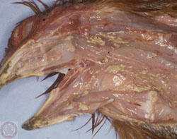
Description:
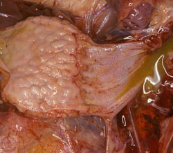
Avian, proventriculus. The proximal mucosa is eroded and covered by a fibrinonecrotic (diphtheritic) membrane.
0215
|
<>
<>
|
|
<>
|
Description:
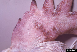
Chicken. The comb is markedly edematous and contains multiple foci of hemorrhage.
Credit: PIADC
0203
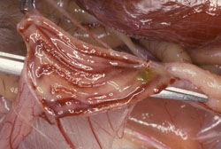
| Description:
Avian, rectum. There are multiple linear mucosal hemorrhages.
0212 |
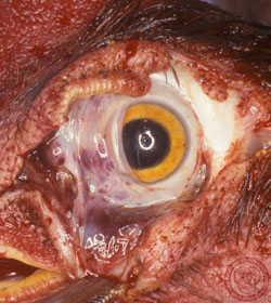
| Description:
Avian, eye. Conjunctival hemorrhage is most severe in the nictitans.
Credit: California Animal Health and Food Safety Laboratory System
0206 |
|
|
|
|
|


No comments:
Post a Comment